Surgical Tooth Extractions
The processBefore a tooth is extracted
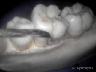
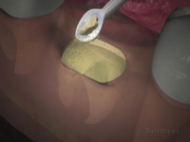
Figure 1: Here a periotome is shown in position in the ligament space on this study skull. Not showing the gums clearly demonstrates the gap that is present between a tooth and the tooth-supporting bone. The tool is inserted as far into the ligament space as possible, to release the majority of the attachment fibers from the tooth.
When you have “invasive” dental procedures like tooth removal done, the dentist will review your health history. If you have replacement joints (for example: knee, hip, etc.), you may be pre-medicated with antibiotics for the procedure. If you have certain types of heart murmurs or replacement heart valves, you may also need to take an antibiotic pre-medication prior to the procedure.
If you take blood thinning medications or drugs that inhibit platelet aggregation, particularly if you take either of the drugs with aspirin, your dentist and/or physician may require you to suspend those medications temporarily to have any oral surgical procedures, including simple tooth removal. This is due to the possibility for prolonged bleeding from the location where the tooth was removed.
If you are anxious about dental procedures, your dentist may recommend sedating you for the procedure. There are several methods of relaxing patients for dental treatment, including oral anti-anxiety pills; inhaled anti-anxiety medication like nitrous oxide; and intravenous anti-anxiety medications. Your dental plan may not pay benefits toward sedation.
The following describes a typical steps involved in surgical tooth removal. Your procedure may vary from the procedure described.
Anesthetic
The area around the tooth to be removed is usually anesthetized with a local anesthetic around the nerve(s) that supply sensation to the tooth. Discomfort from the injection can be minimized by use of a topical numbing gel for a minute or two prior to the injection.
Incision and flap elevation to expose tooth
If the tooth is not visible, or is only partly visible in the mouth, it will be necessary to gently expose the tooth by elevating a surgical flap. An incision is made, and the gum tissues are gently reflected to expose the tooth.
Release periodontal ligament fibers
Teeth are not normally fused to bone. Instead, they have a shock-absorbing ligament that suspends them from the bony tooth socket. The ligament is called the “periodontal ligament”, and the first step in removing a tooth is to release it. This can be done very atraumatically with a thin-bladed instrument called a periotome (Figure 1).
It may be necessary to remove enough bone from around the tooth to allow its removal (full- and partial-bony impactions). This is generally done with a surgical handpiece that is specially designed for the removal of bone.
Safety net
To prevent the tooth (or part of the tooth) from being swallowed or inhaled when it is removed, dentists will often place a “safety net” of gauze in the back of the mouth.
Sectioning of the tooth
Whether or not a surgical access flap is required to expose the tooth, it may be necessary to section it into individual pieces to remove it safely and without trauma. That step would usually be accomplished next. How many pieces the tooth is divided into depends on many factors, including the number and shape of the roots, as well as any nearby anatomical features that may be of concern (like nerves).
Loosening and elevation of the tooth
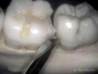
Figure 2: Here an elevator is shown adapted to the back corner of the tooth. Not showing the gums clearly demonstrates that the elevator applies leverage between the tooth and tooth-supporting bone, so as to separate them.
The tooth is (or individual pieces are) loosened (luxated) within the socket by applying leverage with an instrument called an elevator. There are several types of elevators, depending on the shape and size of tooth to be removed, and its location in the mouth. The tooth’s bony housing is somewhat pliable, like a green tree branch, and elevation enlarges the tooth socket enough to remove the tooth (Figure 2). When the tooth has been sectioned, the socket generally isn’t enlarged much, if any.
The tooth fragments are removed in an ordered sequence, that usually involves curved roots being removed last.
Ridge preservation via socket graft (optional)
If the tooth being removed is going to be replaced (for example, with a dental implant or fixed bridge), the dentist may recommend placing bone graft material in the tooth socket to significantly decelerate the bone resorption process and preserve the height and width of the bony ridge at its pre-extraction level. If ridge preservation is not done, the height and width of the bony ridge will immediately begin to deteriorate with the healing process. This procedure is not generally done when teeth are being removed as part of an orthodontic treatment plan.
Placement of an immediate dental implant (optional)
Certain teeth may be candidates for immediate replacement with dental implants. If no acute infection is present (i.e. one that has drainage and swelling), and the bony socket is intact, you and your dentist may plan for immediate placement of the dental implant upon removal of the tooth. A bone graft may be required simultaneously.
Controlling the bleeding
After the tooth is removed, the dentist will apply pressure directly to the tooth socket to minimize any bleeding. There are no major arteries in tooth sockets, so there is usually only a minimal amount of bleeding.
Instructions and care after surgical tooth removal
Your dentist will give you specific instructions, taking into account your unique medical and dental situation. Oral, written, and/or video instructions are helpful. Although they may seem simple at the time of your appointment, questions frequently come up later. Be sure to contact your dentist if you have any questions following this procedure.
Author: Thomas J. Greany, D.D.S. / Editor: Ken Lambrecht
![]()
![]()
![]()
This page was last updated on November 25, 2017.
Related pages on ToothIQ.com